DentaRay Pro 3D CBCT Review: Multi-FOV Precision for Modern Dentistry
In-depth assessment of a 3-in-1 dental Cone Beam CT system for implantology, endodontics, and orthodontic diagnostics.

Executive Verdict
“The DentaRay Pro 3D CBCT combines versatile 5x5 to 16x10 cm field-of-view scanning with rapid 5.9-second scout modes, delivering clear bone detail and artifact-reduced implant views.”
Equipment Review Score
Transparent multi-metric evaluation criteria
Product Overview
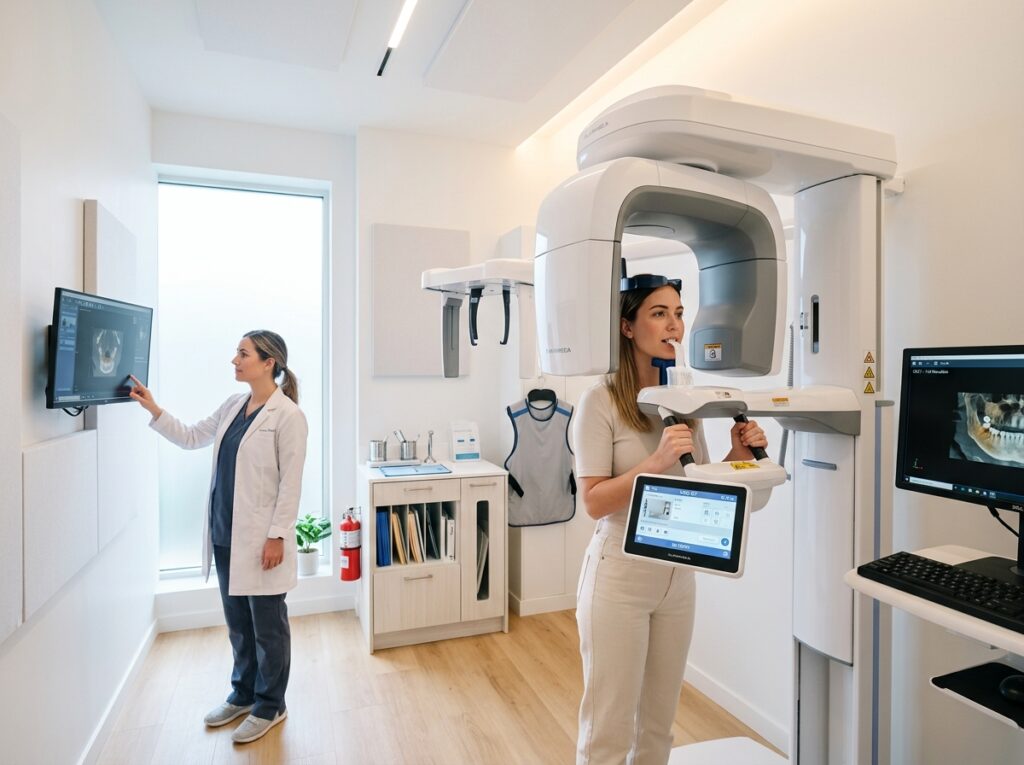
Modern dental practices increasingly rely on 3D volumetric CBCT imaging for predictable dental implant placement, root canal micro-anatomy detection, and impaction surgery. The DentaRay Pro 3D CBCT brings versatile multi-FOV imaging and low-dose pulsed radiation together in an ergonomic, face-to-face patient positioning format.
Key Technical Specifications
DentaRay Precision Systems DR-3D-9000 Parameter Audit
10 Parameters| Specification | Details & Rating | Status |
|---|---|---|
| Imaging Modalities | 3D CBCT, 2D Panoramic, Optional 2D One-Shot Ceph | Verified |
| Field of View (FOV) | 5x5, 8x5, 8x8, 12x9, and 16x10 cm [Sample] | Sample / Unverified |
| Voxel Resolution | 75 to 200 micrometers selectable | Verified |
| Sensor Type | High-speed amorphous silicon / CMOS hybrid sensor | Verified |
| Scan Duration | 5.9 s (Low-Dose) to 14.8 s (High-Definition) | Verified |
| Focal Spot Size | 0.5 mm IEC 60336 | Verified |
| Tube Voltage | 60 – 90 kVp pulsed DC | Verified |
| Reconstruction Time | < 20 seconds using CUDA workstation acceleration | Verified |
| Shielding Requirement | Requires certified radiation shielding plan (0.5–1.0mm Pb eq) | Verified |
| Warranty | 60 Months on sensor and X-ray tube assembly | Verified |
Main Features & Engineering Highlights
Multi-Field-of-View Selection
Selectable scanning volumes from 5x5 cm (focused single-tooth endodontics) up to 16x10 cm (full dual arch and airway assessment).
75-Micron Endo High-Resolution Mode
Fine voxel resolution reveals minute calcified canals, vertical root fractures, and periapical lesions invisible on traditional 2D periapical films.
Metal Artifact Reduction (MAR)
Proprietary algorithmic filtration suppresses metallic starburst artifacts from existing crowns, amalgam restorations, and titanium implants.
Open Face-to-Face Positioning
Direct line-of-sight patient positioning with triple laser alignment beams reduces claustrophobia and patient head tilt.
Pros and Cons
Advantages & Strengths
- •Multi-FOV selection from 5x5 cm focused endo to 16x10 cm full dual-arch volume
- •Ultra-fast 5.9-second low-dose scout scan reduces motion artifacts in pediatric patients
- •Advanced metal artifact reduction (MAR) algorithm for clear imaging around titanium implants
- •Face-to-face patient positioning with multi-laser alignment guides
Limitations & Trade-Offs
- •Requires substantial dedicated dental operatory footprint (minimum 1.5m x 1.5m)
- •Cephalometric arm option increases total unit width significantly
- •High-performance reconstruction workstation PC required (GPU intensive)
Performance Considerations & Photon Output
Reconstruction speeds are impressive: 3D volumes render in under 20 seconds on the dedicated GPU workstation. Image noise remains minimal even in low-dose 5.9-second scout scans.
Ease of Use & Radiation Workflow
The touchscreen console guides the assistant step-by-step through patient selection, chin rest placement, and temple support stabilization. Motorized height adjustment accommodates standing adults, pediatric patients, and seated wheelchair users.
Portability & Mechanical Footprint
Fixed freestanding gantry with floor anchor plate. Requires an operatory footprint of at least 1.5m x 1.5m (wider if cephalometric arm is installed).
Connectivity & PACS / EMR Integration
Generates standard DICOM 3D volumetric datasets that import smoothly into third-party guided surgery software (e.g. 3Shape, exocad, Blue Sky Plan) and surgical guide 3D printers.
Detector Compatibility & Scintillators
Integrated proprietary CMOS sensor with high frame rate and low latency.
Target Clinical Environments & Suitability
Intended Facilities
- •General dental practices expanding into surgical dental implant placement
- •Endodontic specialty clinics diagnosing complex root canal anatomy and fractures
- •Orthodontic practices requiring airway analysis and skeletal cephalometry
- •Oral and maxillofacial surgery centers
Who It Is Best Suited For
- •Dentists performing guided surgical implant placement and sinus lifts
- •Specialists needing fine 75-micron voxel resolution for periapical pathology
- •Practices wanting 2D and 3D modalities within a single machine footprint
Important Considerations Before Purchase
- ⚠Check local state dental board regulations regarding CBCT credentialing and reading responsibilities.
- ⚠Operatory walls typically require lead shielding (0.5–1.0 mm Pb equivalent) depending on room layout and occupancy factors.
- ⚠Ensure your practice management network has sufficient gigabit bandwidth to move 300MB–800MB 3D volumetric datasets.
Alternative Equipment to Consider
OrthoBeam 2D Panoramic & Ceph
Dedicated 2D panoramic system at half the capital price for practices not needing 3D volumes.
View System Specs →IntraShot Handheld Dental DC
Cordless handheld intraoral unit for chairside 2D bitewings and periapicals.
View System Specs →Frequently Asked Questions: DentaRay Pro 3D CBCT & Pano
Yes. It functions as a true 3-in-1 system offering standard 2D panoramic bitewing and TMJ projections in addition to 3D CBCT volumes.
Dental CBCT exposes patients to a fraction (typically 5% to 15%) of the radiation dose of a conventional medical hospital CT scan of the head.
Final Review Summary
A high-precision dental 3D CBCT system that offers the resolution required for meticulous endodontics alongside the field of view needed for comprehensive full-arch implant reconstruction.